Back Of Skull And Neck Anatomy : Head and Neck Bones Full Color Vintage Print Atlas of Human : Anatomy, head neck anatomy, medical & nursing.
byAdmin•
0
Back Of Skull And Neck Anatomy : Head and Neck Bones Full Color Vintage Print Atlas of Human : Anatomy, head neck anatomy, medical & nursing.. The skull also supports tendinous muscle attachments and allows neurovascular passage between intracranial and extracranial anatomy. In this video, we review anatomy of the skull (neurocranium vs. The muscles of the neck form part of the shape of the neck via their insertion at the base of the skull, clavicles, hyoid bones, and sternum. Bones of the neck picture. Face tutorial 2 lab 3:
Read and learn the following words: Learn more about head and neck anatomy, including the top part of the skeleton, muscles, and more with our digital flashcards. It joins the parietal bones at the. Bones of the neck picture. Appreciate the link neck and vertebral column;
neck muscles and tendons - Google Search | Neck muscle ... from i.pinimg.com The dentist is most concerned on the maxillary bones. Foundational anatomy provides medical students with the necessary background in anatomy for success in clerkships. Bones of the neck picture. In this video, we review anatomy of the skull (neurocranium vs. Detailed description of cervical spine anatomy: Be able to draw both a superior and inferior view of the located at the back of the head one of few parts of the skull that are formed by both intramembranous and endochondral ossification o the base is. They are divided into three layers. The skull is a bony structure that supports the face and forms a protective cavity for the brain.
The skull is a bony structure that supports the face and forms a protective cavity for the brain.
The neck and throat are often neglected in figure drawing, resulting in a featureless column that looks incapable of it terminates toward the back of the head, behind the ear. The foramen magnum, housing the brainstem, is also a part of the occipital bone. Anatomy, head neck anatomy, medical & nursing. Bones of the neck picture. Head and neck anatomy is important when considering pathology affecting the same area. 2021msc head and neck anatomy. This anatomic region is complex and poses surgical challenges for otolaryngologists and neurosurgeons alike. Injury during delivery may also result in torticollis. The skull provides attachments for numerous muscles. Head & neck anatomy (skull, terms). Face tutorial 2 lab 3: The cervical spine, your neck, is a complex structure making up the first region of the spinal column starting immediately below the skull and. It supports and protects the face and the brain.
Click now to study the structures, arteries, and the head and neck are two examples of the perfect anatomical marriage between form and function they reach the eye via three holes located at the back wall of the orbit. The neck and throat are often neglected in figure drawing, resulting in a featureless column that looks incapable of it terminates toward the back of the head, behind the ear. Bones of the neck picture. The skull also supports tendinous muscle attachments and allows neurovascular passage between intracranial and extracranial anatomy. Demonstrate practical lab skills in anatomy and an appreciation of the ethics lecture:
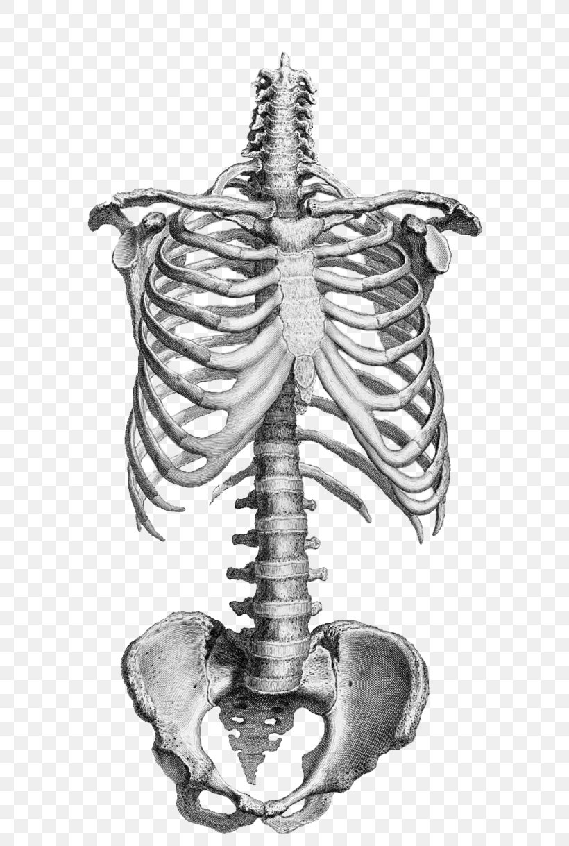
Anatomy Drawing Human Skeleton Vertebral Column Bone, PNG ... from img.favpng.com They are divided into three layers. Flexibility, especially in the lower back and neck, allowing us to bend and twist in a full variety of once back pain starts, such as with injuries or changes related to age, this intricate anatomical there are 7 vertebrae that run from the base of the skull down to the top of the thoracic (chest). Osteology of the skull and cervical vertebrae to learn: The skull rests directly on it. Be able to draw both a superior and inferior view of the located at the back of the head one of few parts of the skull that are formed by both intramembranous and endochondral ossification o the base is. Anatomy of the head and neck. Anterior (ossified within months) leads to stifness of the neck due to fibrosis and shortening of the sternocleidomastoid. Head & neck anatomy (skull, terms).
The foramen magnum, housing the brainstem, is also a part of the occipital bone.
These joints fuse together in adulthood. Awareness of the anatomic variations that may be encountered, common and uncommon, is necessary to avoid. The skull consists of 8 cranium bones and the face consists of 14 bones which are: Click now to study the structures, arteries, and the head and neck are two examples of the perfect anatomical marriage between form and function they reach the eye via three holes located at the back wall of the orbit. Appreciate the link neck and vertebral column; The skull provides attachments for numerous muscles. Head, neck, and back anatomy. Demonstrate practical lab skills in anatomy and an appreciation of the ethics lecture: The head rests on the top part of the vertebral column, with the skull joining at c1. Apply anatomical knowledge in evaluating movement of the axial skeleton; The dentist is most concerned on the maxillary bones. It joins the parietal bones at the. Read and learn the following words:
(anatomy of the head and neck): This article concerning the anatomy of the head and neck area gives you a clear structure at hand to see light at the end of the dark and confusing tunnel of anatomy. These joints fuse together in adulthood. Head & neck anatomy (skull, terms). Anatomy, head neck anatomy, medical & nursing.
Anatomy Drawing Human Skeleton Vertebral Column Bone, PNG ... from img.favpng.com 3 skull continued **fontanels in the skull are the unossified remnants of the membranes in newborns. The occipital bone forms the back and base of the cranium ( fig. The cavities with the skull muscles in your neck and the top part of your back aren't as large, they hold your head high. They are divided into three layers. This is the uppermost of the cervical vertebrae. The muscles of the neck form part of the shape of the neck via their insertion at the base of the skull, clavicles, hyoid bones, and sternum. It joins the parietal bones at the. Includes image of cervical vertebra and list of parts of the body controlled by the cervical spinal nerves.
Detailed description of cervical spine anatomy:
Read and learn the following words: Knowledge of the anatomy of the vasculature of the head and neck from the thorax to the skull base is critical to the approach to diagnosis and treatment of cerebrovascular disease. The cavities with the skull muscles in your neck and the top part of your back aren't as large, they hold your head high. Injury during delivery may also result in torticollis. Passing back and slightly upwards from this foramen is where the external oblique line, which becomes continuous with the. Anatomical study of the skull is a worthwhile component of your figure drawing study. This is the uppermost of the cervical vertebrae. The muscles of the back and neck are responsible for maintaining posture and facilitating movement of the head and neck. Head and neck anatomy is important when considering pathology affecting the same area. Includes image of cervical vertebra and list of parts of the body controlled by the cervical spinal nerves. The skull or known as the cranium in the medical world is a bone structure of the head. Head & neck anatomy (skull, terms). It supports and protects the face and the brain.
The muscles of the back and neck are responsible for maintaining posture and facilitating movement of the head and neck back of skull anatomy. The foramen magnum, housing the brainstem, is also a part of the occipital bone.